Section 1: Basic Characterization
Section 2: Pluripotency and the Undifferentiated State
Section 3: Genomic Characterization
Section 4: Stem Cell-based Model Systems
Appendix 1: Recommended Standard Characterization of Stem Cells
Appendix 2: Nomenclature Criteria
Appendix 3: Cell Culture Hygiene Practices
Appendix 5: Assessment of Methods for Genetic Analysis
Appendix 6: Reporting Practices for Publishing Results with Human Pluripotent and Tissue Stem
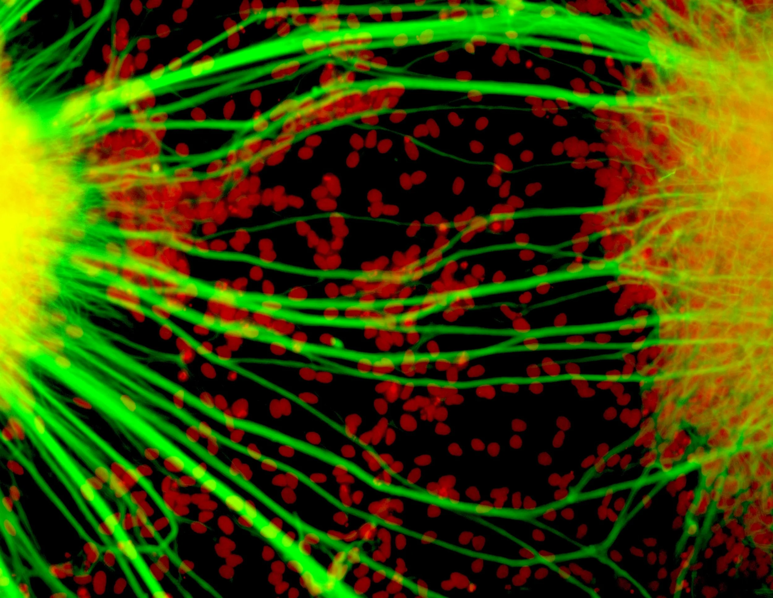
Appendix 4: Markers for the Identification of Undifferentiated hPSCs and Monitoring Multi-Lineage Differentiation
Few, if any, gene transcripts, proteins, or oligosaccharides are uniquely expressed by a single cell type so that identification and monitoring of undifferentiated hPSCs, or of cells corresponding to ectoderm, mesoderm or endoderm require the use of multiple markers. Building on a set of cell surface antigens previously defined and characterized in human embryonal carcinoma cells (Andrews et al., 1996), the International Stem Cell Initiative identified a set of cell surface markers (Table A4.1) that are typically expressed by undifferentiated human ES cells (The International Stem Cell Initiative, 2007). The International Stem Cell Initiative also used transcriptome analysis of a panel of genes (Table A4.2, Table A4.3) to monitor undifferentiated human hPSCs and their differentiation into derivatives corresponding to ectoderm, mesoderm and endoderm. A similar panel of genes was used in the study by Bock et al (2011), which was then used by Tsankov et al (2015) to quantify differentiation to the three germ layers.
Table A4.1. Cell surface marker antigens for undifferentiated human ES cells

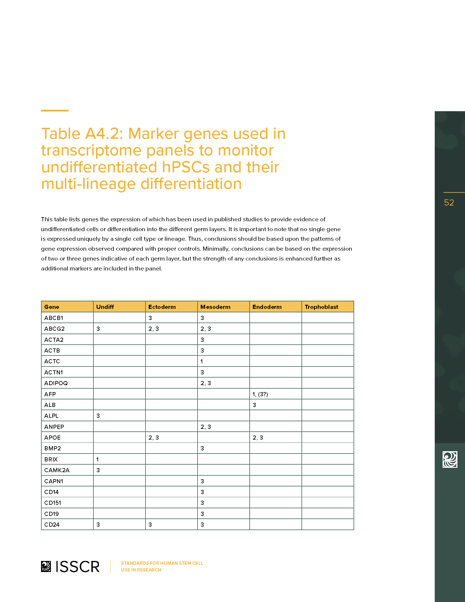
Table A4.2: Marker genes used in transcriptome panels to monitor undifferentiated hPSCs and their multi-lineage differentiation
This table lists genes the expression of which has been used in published studies to provide evidence of undifferentiated cells or differentiation into the different germ layers. It is important to note that no single gene is expressed uniquely by a single cell type or lineage. Thus, conclusions should be based upon the patterns of gene expression observed compared with proper controls. Minimally, conclusions can be based on the expression of two or three genes indicative of each germ layer, but the strength of any conclusions is enhanced further as additional markers are included in the panel.
1) Characterization of human embryonic stem cell lines by the International Stem Cell Initiative (The International Stem Cell Initiative, 2007).
2) Reference maps of hESC and hiPSC variation enable high-throughput characterization of pluripotent cell lines (Bock et al., 2011).
3) Assessment of established techniques to determine developmental and malignant potential of human pluripotent stem cells by the International Stem Cell Initiative (Allison et al., 2018).
Table A4.3: Reduced marker gene set used for embryoid body analysis

Xenograft Tumor Assays
We have not recommended the routine use of xenograft tumor (teratoma) assays to assess pluripotency since in vitro assays can adequately confirm the ability of hPSC to differentiate into cells of the ectoderm, mesoderm, and endoderm lineages. However, xenograft tumor assays may be useful to assess the degree to which the differentiated derivatives of hPSC can undergo maturation and histogenesis. Further, xenograft assays can indicate malignant potential of a cell line, revealed by the presence of undifferentiated pluripotent stem cells, or yolk sac elements and immature neurectoderm, within the tumor (Allison et al., 2018). In addition, xenografts can reveal if patient-derived cultures considered “healthy” contain tumorigenic cells (Georgakapoulos et al., 2020). If xenograft assays are used, full details of the strain of mouse used, the site and method of inoculation and the method of cell preparation should be provided. The histology of tumors should be assessed by a qualified histopathologist using WHO criteria and/or with reference to suitable histology atlases (Damjanov and Andrews, 2016). The term teratoma’ should be reserved for tumors containing tissues corresponding to all three germ layers, but without evidence of undifferentiated stem cells; the term teratocarcinoma should be used for tumors containing tissues corresponding to all three germ layers and undifferentiated stem cells (Damjanov and Andrews, 2007). Traditional histological morphological approaches can be combined with computational approaches, such as the Teratoscore assay (Avior et al., 2015), in which RNA-seq data from teratomas is used to obtain detailed information on their cellular composition, including the presence of undifferentiated stem cells (Allison et al., 2018).
Appropriate antigens for the detection of undifferentiated stem cells and yolk sac elements are listed below.
Undifferentiated stem cells: OCT4, TNFRSF8, TRA-1-60/GCTM2
Yolk sac carcinoma: CDX2, GPC3, AFP, SALL4
Primitive neuroectoderm: PAX6, CD99, CD56, POU5F1, TNFRSF19
The ISSCR's Standards for Human Stem Cell Use in Research are strictly copyrighted by the society. No part of this document may be produced in any form without written permission of The International Society for Stem Cell Research. Contact isscr@isscr.org for more information.
©2026 by The International Society for Stem Cell Research. All rights reserved.