
Building Functional Stem Cell Systems
An ISSCR Webinar featuring ISSCR 2026 Poster Award Winners
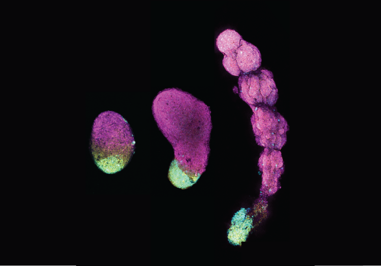
Image: Pranav Saligrama Ramesh, University of Calgary, Canada
Program Description: Discover ways researchers are building functional systems to probe gene regulation, model disease, and engineer organ-level physiology in a variety of pluripotent stem cell systems. Presented by ISSCR 2026 Poster Award Winners, this program is a unique opportunity to learn from emerging leaders and stay at the forefront of stem cell science.
The webinar will take place on 29 September 2026 at 11:00 a.m. - 12:30 p.m. EDT
View when the webinar is happening in your time zone.
Registration for this webinar is open to all audiences.
As an ISSCR member benefit, members are invited to register at no cost.
The registration fee for non-members is $150.00 USD.
Non-members from countries identified by the World Bank as "low- and low-middle income countries"
receive a 50% reduction on registration rates. Eligible countries can be found here.
Registration will close 3 hours prior to the beginning of the webinar
The webinar will be hosted via Zoom. Registrants will receive an email reminder with the webinar link one day, and one hour prior to the event.
Moderators
Kai Kretzschmar, PhD
University of Würzburg, Germany
Poster Award Winners & Presenters
Elodie Limberg
Max Planck Institute for Molecular Genetics, Germany
Presentation Title: HIGH-THROUGHPUT MAPPING OF FUNCTIONAL INTERACTIONS BETWEEN CIS-REGULATORY ELEMENTS AND TRANSCRIPTION FACTORS IN MOUSE EMBRYONIC STEM CELLS
From Edda Schulz’s Lab
-
Regulation of gene expression involves intricate interactions between transcription factors (TFs) and cis-regulatory elements (CREs), where multiple CREs and TFs functionally interact to control a single gene. Yet the specific TF–CRE interactions that control gene expression at endogenous loci remain difficult to dissect. To address this, we have developed a combinatorial approach that integrates CRISPR/Cas9-directed transcription factor perturbation screens with Massively Parallel Reporter Assays (MPRA). The core component is a genomically integrated construct that co-expresses an sgRNA inducing TF repression together with a CRE-driven reporter gene, enabling simultaneous perturbation of TF activity and quantitative measurement of cis-regulatory element activity. This strategy allows us to systematically identify functional TF–CRE interactions by measuring how libraries of regulatory elements respond to altered TF levels. Our efforts also include the evaluation of integration methods such as piggyBac and integrase systems to achieve high efficiency of single-copy integration in mouse embryonic stem cells. We apply this framework to the Xist locus, the master regulator of X-chromosome inactivation in mouse embryonic stem cells. Candidate CREs were selected from previously identified Xist cis-regulatory elements and prioritized using base-resolution contribution scores from ChromBPNet models. Using this system, we systematically map TF–CRE interactions controlling Xist regulation during early differentiation of mouse embryonic stem cells, demonstrating the potential of this approach to dissect regulatory networks at complex genomic loci. In the future, this framework could enable a more systematic and scalable dissection of TF–CRE interactions across diverse genes and cellular contexts.

Holly Poling
Cincinnati Children's Hospital and Medical Center, USA
Presentation Title: ENGINEERING FUNCTIONAL INTESTINE: FED-STATE ADAPTATION OF IPSC-DERIVED TISSUE IN THE FECAL STREAM
-
A long-standing goal of tissue and organoid engineering is the generation of fully functional, transplantable organs capable of integrating with physiological systems. In the intestine, decellularized extracellular matrix scaffolds and organoid-seeded mucosal grafts have demonstrated short-term engraftment and luminal structure formation, but have not achieved sustained physiological integration or functional adaptation in vivo, highlighting the need for alternative strategies that enable long-term, coordinated epithelial and neuromuscular maturation. Here, we present an induced pluripotent stem cell (iPSC)–based confined culture system (CCS) that generates anatomically laminated intestinal tissue and evaluate its capacity to function under true physiological challenge. By establishing luminal continuity between CCS-derived intestine and the host fecal stream, we modeled the transition from a sterile to a fed intestinal state. CCS constructs not only survived this challenge for 12 weeks, but exhibited adaptive remodeling across multiple compartments, including dynamic regulation of epithelial barrier properties, mucin production, and ion transport. Importantly, luminal content exposure enhanced neuromuscular maturation, with elevated non-stimulated contractile amplitudes and increased basal muscle tone compared to sham-operated controls. Together, this work provides proof of concept that iPSC-derived, engineered intestinal tissue can be transplanted, placed into the fecal stream, and withstand physiological demands while adapting as an integrated, organ-level system. This platform establishes a benchmark for functional validation in intestinal tissue engineering and represents a step toward realizing fully engineered organs capable of meeting translational performance criteria.
Pranav Saligrama Ramesh
University of Calgary, Canada
Presentation Title: BALANCING FGF AND WNT SIGNALING ACTIVITY ENABLES REPRODUCIBLE GENERATION OF HUMAN SOMITOIDS
-
Parvalbumin positive (PV+) cortical interneurons play a critical role in regulating cortical network synchrony and γ-band oscillations, and their disruption in schizophrenia is thought to contribute to the cognitive symptoms of the disorder. Although defects in cortical interneurons have long been reported in schizophrenia patients, how such defects emerge during development remains poorly understood. An in vitro human system to study PV+ cortical interneurons in the context of schizophrenia and other psychiatric disorders would provide an invaluable resource for both mechanistic and translational studies; however, these cells have been notoriously difficult to derive from human pluripotent stem cells, likely owing to their protracted maturation times in vivo. Here, we have developed a forebrain assembloid system which supports the development and functional maturation of PV+ cortical interneurons. We show that cortical interneurons in forebrain assembloids upregulate parvalbumin in as little as 4 months in vitro, express the expected molecular profile, display the characteristic morphology of PV+ basket cells, and the distinct electrophysiology of fast-spiking cortical interneurons. Further, forebrain assembloids containing PV+ cortical interneurons generate γ-band oscillations, a property consistently disrupted in patients with schizophrenia. To assess the development of cortical interneurons in the context of psychiatric disease, we engineered a set of isogenic schizophrenia-associated structural variants in human embryonic stem cells using CRISPR. We found that deletion of 22q11.2 led to disrupted tangential migration in developing cortical interneurons, which we have linked to disrupted mitochondrial function. Further, we observe evidence of disrupted cortical network activity, including decreased γ-band oscillations, in multiple schizophrenia-associated structural variants. These findings provide mechanistic insight into how deficits in cortical interneuron development may contribute to schizophrenia and provide a robust, reliable platform for the derivation and study of PV+ cortical interneurons in human disease.
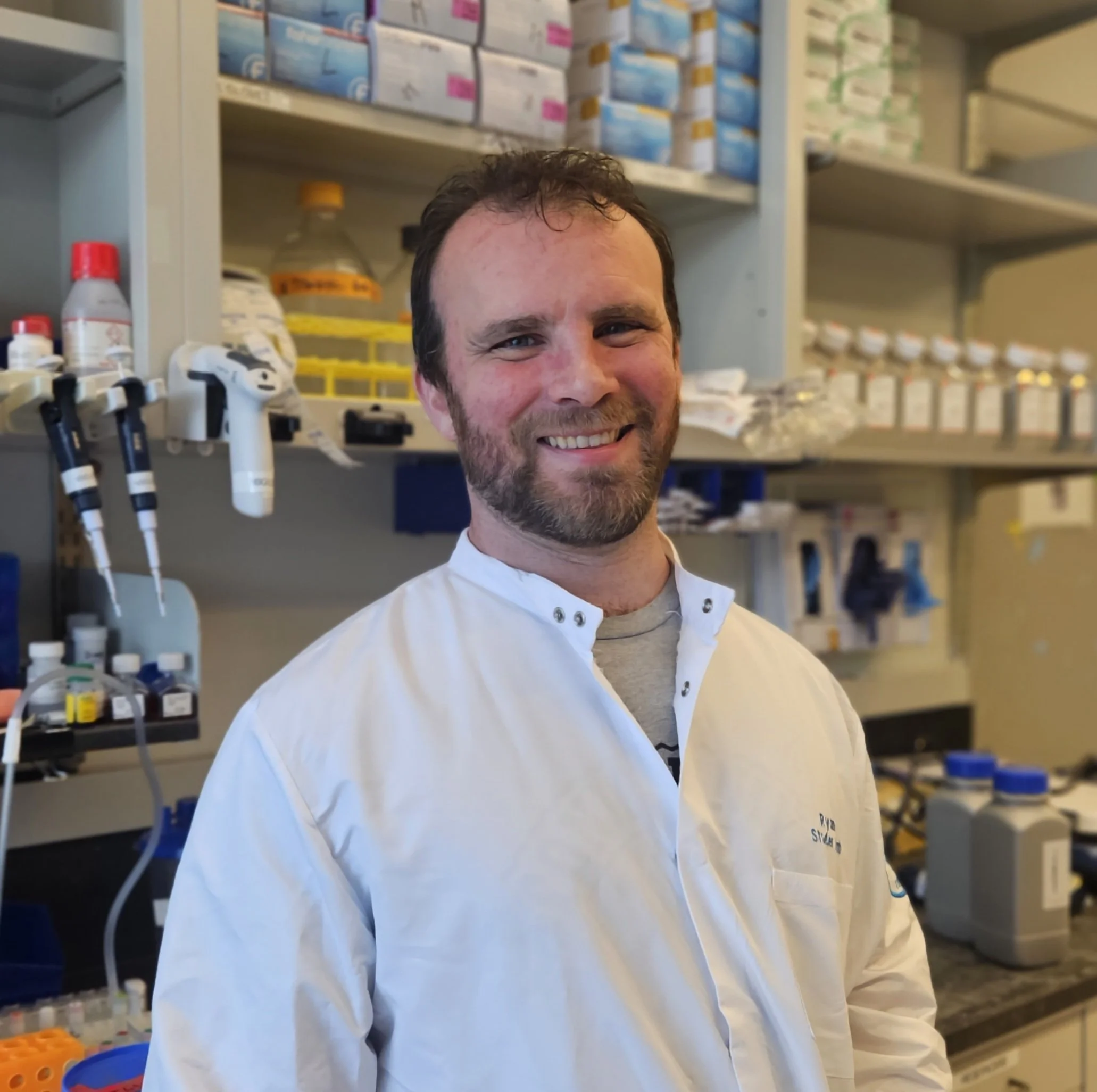
Ryan Walsh, PhD
Memorial Sloan Kettering Cancer Center, USA
Presentation Title: MODELING SCHIZOPHRENIA IN FOREBRAIN ASSEMBLOIDS CONTAINING FUNCTIONAL PV+ CORTICAL INTERNEURONS
From Lorenz Studer’s Lab
-
Human pluripotent stem cell (hPSC)-derived models of somitogenesis (or “somitoids”) demonstrate a remarkable ability to self-organize in response to morphogen signaling, undergo axial elongation, and show periodic somite formation. Somitoids hold great potential for studying the fundamental principles that shape the vertebrate body plan. Yet, current model systems vary widely in their differentiation strategies and exogenous morphogen cues provided to generate somites. This limits generalizability across different hPSC lines and obscures the core signaling network that drives somitogenesis. To systematically optimize somitoid generation, we employed an endogenous MEOX1 reporter system in human embryonic stem cells (hESCs) to track somite fate commitment in real time. We show that FGF and WNT signaling cues have opposing effects on the onset of spatial MEOX1 expression, beginning approximately 3.5 days post-aggregation and preceding periodic somite segmentation. Striking a precise balance between the two signaling pathways ensures proper fate commitment, which is both predictive and required for proper segmentation clock activity, axial elongation, and somite budding in somitoids. Unbiased single-cell RNA sequencing using Parse technology demonstrated that somitoids faithfully recapitulate the cellular composition and transcriptional trajectories of vertebrate somitogenesis. Comparative analysis further revealed remarkable similarities between somitoids and Carnegie stage 12-16 human embryos. Modulating the FGF and WNT signaling balance according to this defined logic was sufficient to overcome line-to-line variability, allowing reproducible generation of somitoids across seven parental hESC and hiPSC lines. Altogether, we anticipate that this defined signaling framework will enable a robust modelling of early human development and help better investigate idiopathic congenital disorders in vitro.